Every day is an adventure with primary care cases. From allergies to rashes and everything in between, providers are sharing their most interesting medical cases in primary care to Figure 1. Here are five cases attracting a lot of buzz.
#5. A Surprising Find
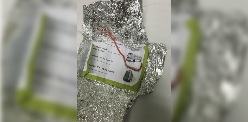
A parent and their 6-year-old child came in for care and brought a ball of tinfoil with them. The registered nurse who treated the child shared the surprise found inside the foil with the Figure 1 community, “Was expecting to unwrap tin foil and see pin worms …”
What is that in the tinfoil? View the case and join the conversation
#4. Persistent Rash
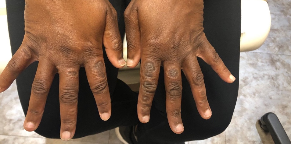
The physician assistant who shared this interesting primary care case noted, “Early 40s female with history of hypertension. Works in a prison. For over six months has this rash on her hands. Frequently uses sanitizer and latex-based gloves while working. Otherwise healthy. Hands are itchy … Did not do DIF but asked dermatopathologist if they recommend based on what they see.”
Ideas range from a fungal infection to Gottron papules, and contact dermatitis.
What do you think is the cause of this rash? View the case and find out if you’re right
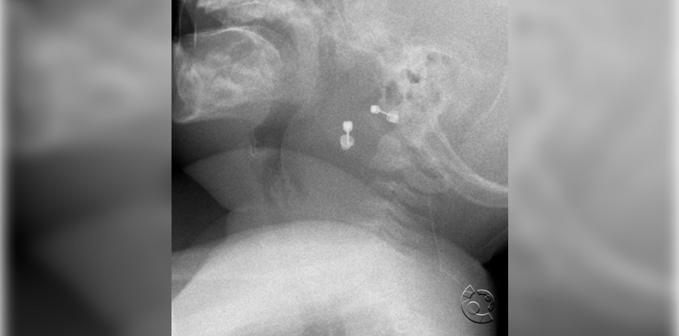
#3. Bulging and Enlargement of the Retropharyngeal Soft Tissue
In this case, we see an infant’s lateral radiograph. While you might first be drawn to the two visible earrings, they are not the troubling part of the image. In The Differential: Primary Care newsletter, Dr. Harrison Hayward noted, “The prevertebral retropharyngeal space is quite wide, concerning for retropharyngeal abscess. A contrast CT provides the definitive diagnosis, but angering this child with an IV stick might compromise their airway.”
What’s your next step? View the case and share your comments
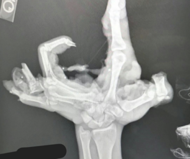
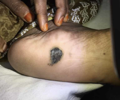
#2. A Growing Lesion on the Leg
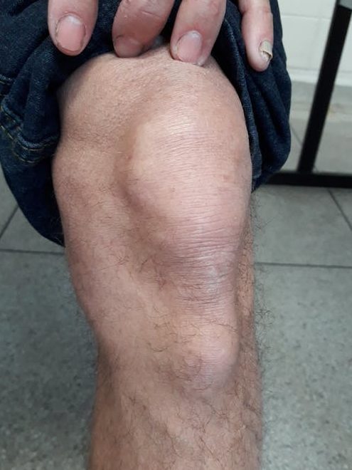
This interesting primary care case describes a 69-year-old male patient who presented with this lesion in his leg that has been growing in size over the last 12 months. According to the Figure 1 member who shared the case, the lesion is painless, hard, motionless, and with no previous trauma. An X-ray is the next step.
One commenter noted, “The fact that it is hard excludes bursitis. Osgood-Schlatter appears to be the best fit, clinically. An X-ray should clinch the dx, revealing a prominent tibial plateau.” But another countered with, “Would seriously doubt Osgood-Schlatter in this patient. Physis has been sealed for the better part of five decades and without a possible antecedent injury it is not likely. Osteochondroma of the tibial tuberosity more likely, or a hardened bursitis, or rheumatoid nodules … Would be interested to see the X-ray when it comes back.”
What’s your differential? View the case and weigh in
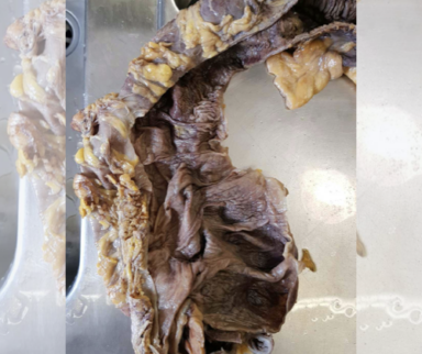
#1. Intermittent Chest Pain and Dyspnea
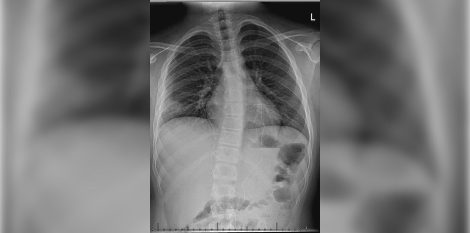
In this case, we see the scan of an 11-year-old boy after three weeks of intermittent chest pain and dyspnea, but no fever or cough. According to the Figure 1 member who shared this case, “Diminished breathing sounds lower right. CRP < 1, normal blood gas, normal ECG. What’s next? Antibiotics … or chest CT or CT angio (PE)?”
The case prompts a dialogue around the risks and benefits of further scans for young patients.
Do you proceed with additional scanning or take a different approach? See what the care team decided
Published January 25, 2022
Join the Conversation
Register for Figure 1 and be part of a global community of healthcare professionals gaining medical knowledge, securely sharing real patient cases, and improving outcomes.