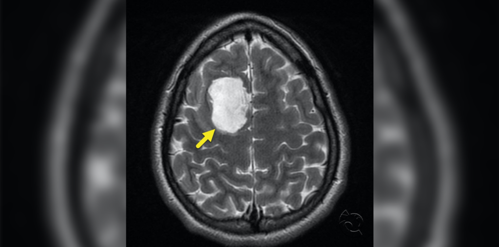
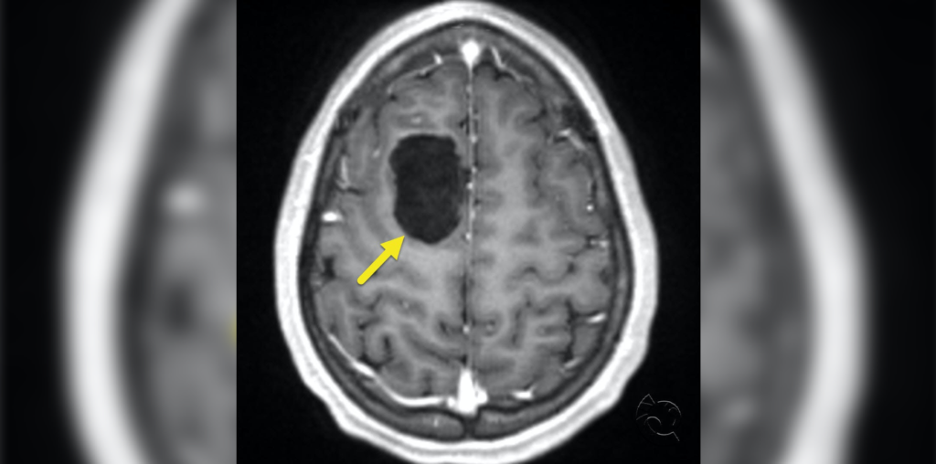
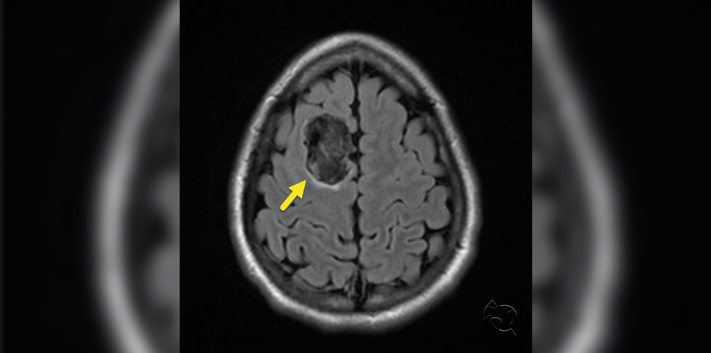
A 15-year-old undergoes brain MRI after a mild traumatic brain injury. Axial T2 (image 1), T1 postcontrast (image 2), and FLAIR (image 3) sequences show a cystic lesion involving the right superior frontal gyrus with no concerning edema or mass effect.
The lesion has mild heterogeneous signal on FLAIR and shows restricted diffusion and no contrast enhancement. These features are characteristic of an epidermoid cyst. Epidermoid cysts most commonly occur at the cerebellar pontine angle or the region of the fourth ventricle, but they can also occur supratentorially. They are usually very slow growing.
FLAIR and DWI sequences are useful for differentiation of these lesions from arachnoid cysts due to their heterogeneous FLAIR signal, higher than cerebrospinal fluid, and abnormal diffusion restriction with T2 shine-through. Surgical excision is the treatment of choice if symptomatic, but no treatment is indicated in an asymptomatic patient.
Further Reading
Intraparenchymal Epidermoid Cyst: Proper Surgical Management may Lead to Satisfactory Outcome
Intrinsic Brainstem Epidermoid Cyst. Case Report and Review of the Literature
Reference
Dutt SN, Mirza S, Chavda SV, Irving RM. Radiologic differentiation of intracranial epidermoids from arachnoid cysts. Otol Neurotol 2002;23(1):84-92.

Aravind Ganesh, MD, DPhil(Oxon), FRCPC
Vascular and Cognitive Neurologist and Assistant Professor, University of Calgary Department of Clinical Neurosciences
Published February 17, 2022
Want more clinical cases?
Join Figure 1 for free and start securely collaborating with other verified healthcare professionals on more than 100,000 real-world medical cases just like this one.