A 76-year-old undergoes cardiac surgery, during which they develop ventricular fibrillation.
After 15 minutes of direct cardiac massage, they are put on extracorporeal membrane oxygenation (ECMO). Following a return to sinus rhythm, they are in a deep coma.
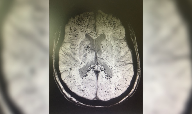
Brain MRI shows multiple small hypointense lesions on susceptibility weighted imaging (SWI) sequences. These lesions are cerebral microbleeds, which can often be seen after ECMO in both adults and children, and are thought to reflect vascular damage from gaseous emboli.
Distinct patterns of microbleeds can also be seen in the cerebral white matter in other critically ill patients and may relate to hypoxemia, as they are similar in appearance to those described with high-altitude exposure.
Case series have suggested that patients with ECMO-associated microbleeds without other imaging abnormalities can have a favorable outcome despite the worrisome imaging appearance, and indeed, this patient improves markedly over a few days.
Further Reading
Figure 1 Medical Case: Cardiac Surgery After Iatrogenic Tamponade
Diffuse Cerebral Microbleeds After Extracorporeal Membrane Oxygenation Support
Cerebral Microbleeds After Use of Extracorporeal Membrane Oxygenation in Children
References
Fanou EM, Coutinho JM, Shannon P, et al. Critical illness-associated cerebral microbleeds. Stroke 2017;48(4):1085-1087.

Aravind Ganesh, MD, DPhil(Oxon), FRCPC
Vascular and Cognitive Neurologist and Assistant Professor, University of Calgary Department of Clinical Neurosciences
Want more clinical cases?
Join Figure 1 for free and start securely collaborating with other verified healthcare professionals on more than 100,000 real-world medical cases just like this one.