Chondrosarcoma, a group of malignant bone tumors, is diagnosed in fewer than 1,000 patients a year in the United States. Rare diseases like chondrosarcoma underline the value of the Figure 1 platform: It’s a place where millions of healthcare professionals around the world can see and discuss detailed real-world cases they may never otherwise encounter in their everyday practices.
Here are some of the top chondrosarcoma cases on Figure 1 as shared and discussed by oncologists, orthopedic surgeons, and radiologists:
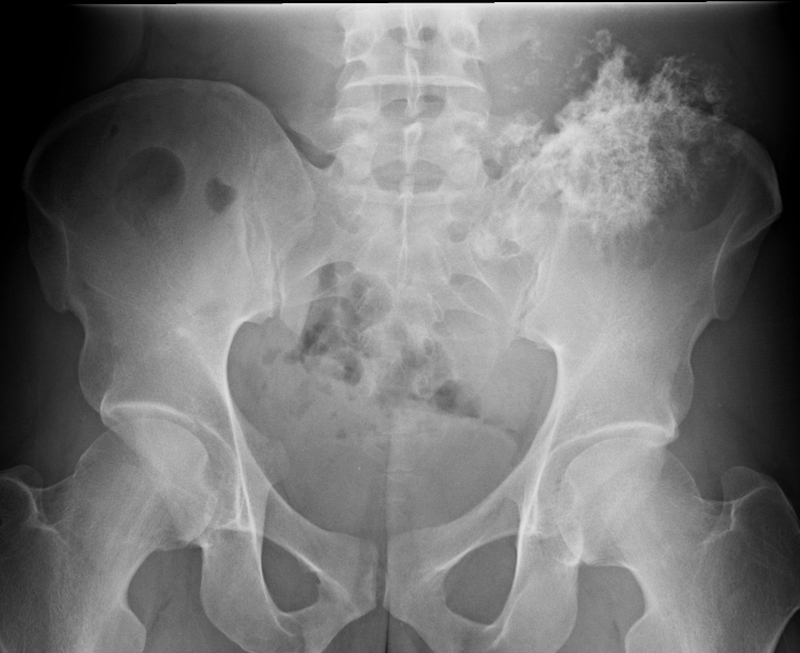
1. Multiple Osteochondromas Arising From the Pelvis
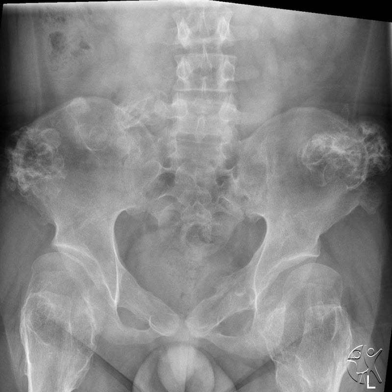
This AP X-ray of the pelvis shared by the Radiology and Nuclear Medicine department of Cincinnati Children’s Hospital shows multiple osteochondromas arising from the pelvis.
“Many of the lesions arising from the pelvis are pedunculated while the lesions of the femurs are more sessile. Patients with multiple hereditary exostoses (bone spurs) are at higher risk of developing chondrosarcoma.” – Cincinnati Children’s Hospital
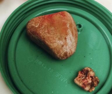
2. Chondrosarcoma treated with limb salvage surgery
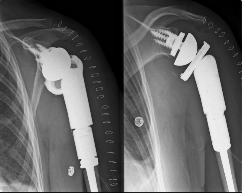
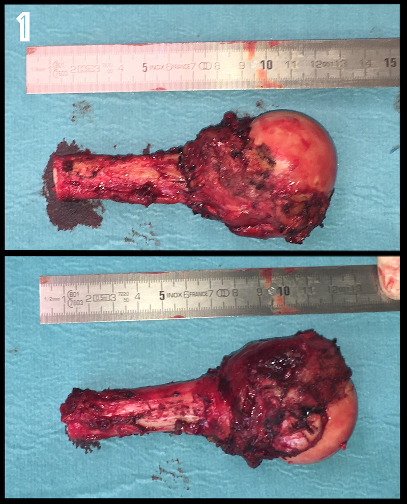
“Here’s a case of a 44-year-old woman with a pathological fracture of the humerus. Medullary chondrosarcoma can destroy and weaken the bone from the inside so much that remaining cortices aren’t strong enough for any kind of fixation. After studying her case with CT and MRI, the lead surgeon and his team decided to go for a reversed shoulder megaprosthesis. Rotator cuff tendons were reattached on the prosthesis.” – Orthopedic Surgeon
Included are images of the resected bone:
Read a detailed explanation of the case here
Learn more about treating sarcoma with radiation therapy from Cancer, a journal of the American Cancer Society, on Figure 1
3. The Initial Diagnosis of Chondrosarcoma
“A 30-year-old male who complained of a painful mass above his left hip. Pain started 3 months prior to this X-ray according to the patient. This was the result a few months later: huge chondrosarcoma arising from the iliac bone.” – Orthopedic Surgeon
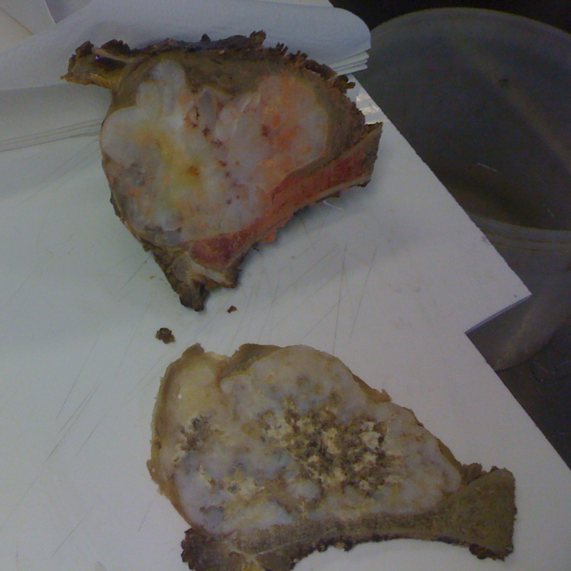
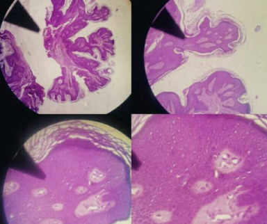
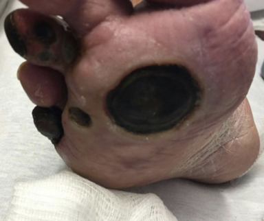
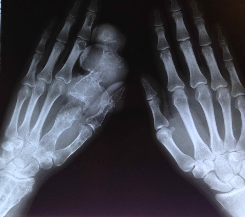
4. Ollier Disease and Chondrosarcoma
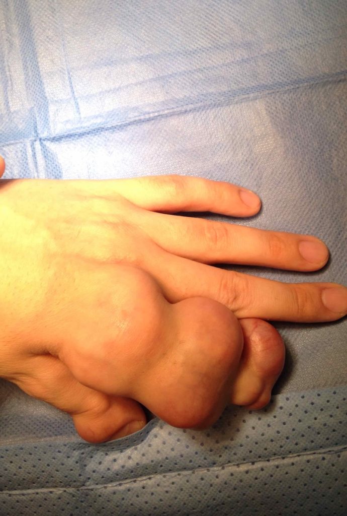
Ollier disease is a rare form of skeletal dysplasia that is characterized by benign tumors of the cartilage called enchondromas. Rarely, these tumors may be complicated by malignant transformation to chondrosarcoma. In this case shared by an orthopedic surgeon, a patient with Ollier disease developed a chondrosarcoma, necessitating amputation of the affected digits.
Published October 20, 2021
Join the Conversation
Register for Figure 1 and be part of a global community of healthcare professionals gaining medical knowledge, securely sharing real patient cases, and improving outcomes.